EN
Translate:
Many eye diseases develop silently, without noticeable symptoms. The American Academy of Ophthalmology (AAO) has cited research showing that 63% of people found to have eye disease were unaware of their condition.¹
Children are not immune to eye health problems. Research has found retinal abnormalities in young adults with myopia who had no symptoms. A 2024 study published in Ophthalmology, the AAO's journal, identified peripheral retinal abnormalities in 16.1% of eyes, including retinal breaks or detachments in 4.7%.²
In our own practice, we have discovered retinal holes in children as young as seven years old, including cases requiring laser treatment—even though they had no noticeable symptoms.
These findings remind us that good vision does not necessarily mean healthy eyes.
This is why we believe comprehensive eye examinations and advanced diagnostic technology are so important. Technologies such as Optomap® ultra-widefield retinal imaging help our doctors examine areas of the retina that may be difficult to assess through conventional methods alone.
By combining advanced technology, clinical experience and patient education, we strive to detect eye problems early, monitor changes over time and protect your vision throughout life.
Our goal is not simply to help you see clearly, but to help keep your eyes healthy for life.
References
1. American Academy of Ophthalmology. Comprehensive Adult Medical Eye Evaluation Preferred Practice Pattern. 2020.
2. Ultra-Widefield–Guided Swept-Source OCT Findings of Peripheral Vitreoretinal Abnormality in Young Myopes. Ophthalmology. 2024. Study of asymptomatic myopic adults aged 18–42.


Most parents would never imagine that a child with no visual complaints could have a retinal hole.
Yet in our practice, we have discovered retinal holes in children as young as seven years old, including cases requiring laser treatment.
These children had no noticeable symptoms.
Good vision does not always mean a healthy retina.
*Actual Optomap image of a seven-year-old patient from our practice.
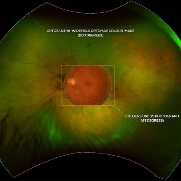
Since 2008, our practice has used Optomap® ultra-widefield scanning laser technology to help detect retinal abnormalities, often before patients experience any symptoms.

Optomap® retinal scan and traditional retinal examinations provide complementary information. Combining advanced imaging with careful clinical examination helps us assess retinal health more thoroughly.
During a traditional retinal examination, an ophthalmoscope allows the doctor to view a limited area of the retina at one time. Patients are therefore asked to look in different directions so the doctor can examine more of the retina.
Conventional retinal photography typically captures approximately 35–45° of the retina in a single photograph. In contrast, Optomap uses scanning laser technology to capture up to 200° of the retina in a single scan, in less than 0.4 seconds, providing a much wider view of the peripheral retina.
This quick, widefield scan can be particularly helpful when examining young children or patients who have difficulty keeping their eyes steady during an examination.
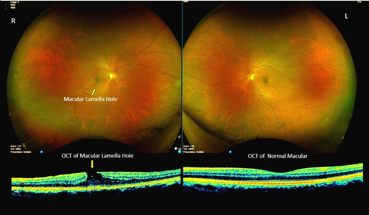
Optomap provides a wide view of the retina, but some eye diseases cause changes within the retinal layers that may not be obvious on a retinal image.
Optical Coherence Tomography (OCT) uses light to create highly detailed, cross-sectional images of the retina, allowing our doctors to examine its individual layers.
Combining Optomap with OCT helps us see both the broader retinal picture and the detailed structures beneath the surface.
Can you see the difference?
The retinal images above look remarkably similar, but OCT reveals a lamellar macular hole in the right eye, while the left macula has a normal structure.

OCT Macular Scan can help detect and monitor changes associated with age-related macular degeneration (AMD), diabetic macular edema, myopic macular degeneration and epiretinal membranes, sometimes before patients notice changes in their vision.
The example above shows how OCT reveals retinal swelling associated with diabetic macular edema that may be difficult to appreciate from retinal images alone. Notice also the small retinal hemorrhages visible on the Optomap image.
OCT measures the thickness of the retinal nerve fibre layer (RNFL) and ganglion cell complex (GCC), helping our doctors detect and monitor changes associated with glaucoma.
These detailed measurements may reveal early structural changes before vision loss can be detected on a visual field test. Repeated OCT scans also provide objective measurements to help monitor glaucoma progression over time. OCT may also help identify optic nerve swelling associated with certain neurological conditions, including increased intracranial pressure, which may require further medical investigation.
OCT Corneal pachymetry measures corneal thickness, sometimes at multiple points across the cornea. These measurements help assess certain corneal conditions, evaluate patients undergoing refractive surgery, and interpret eye pressure measurements when assessing glaucoma risk.
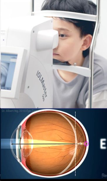
Most people don’t realize that an excessively long eye increases the lifetime risk of serious eye diseases.
Axial length is also an important indicator of myopia progression and, together with other clinical findings, helps identify children at risk of faster progression who may benefit from more intensive myopia management.
By monitoring
Most people don’t realize that an excessively long eye increases the lifetime risk of serious eye diseases.
Axial length is also an important indicator of myopia progression and, together with other clinical findings, helps identify children at risk of faster progression who may benefit from more intensive myopia management.
By monitoring eye growth annually, our doctors can assess myopia progression and the effectiveness of treatment.
Our advanced Medmont Meridia® corneal topographer analyzes up to 102,000 data points to create a detailed map of the cornea's shape and curvature.
This information helps our doctors precisely design and fit AOK Eye Braces® (Ortho-K) for optimal vision correction and myopia control. It also helps detect early corneal abnormalities, includi
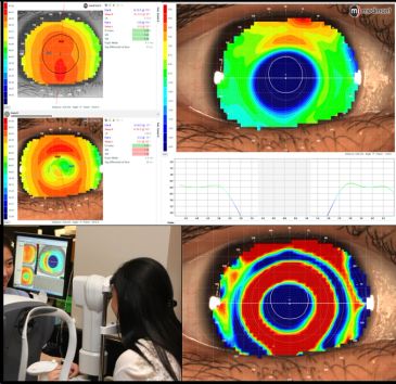
Our advanced Medmont Meridia® corneal topographer analyzes up to 102,000 data points to create a detailed map of the cornea's shape and curvature.
This information helps our doctors precisely design and fit AOK Eye Braces® (Ortho-K) for optimal vision correction and myopia control. It also helps detect early corneal abnormalities, including signs of keratoconus, and assess corneal suitability for laser vision correction and cataract surgery planning.
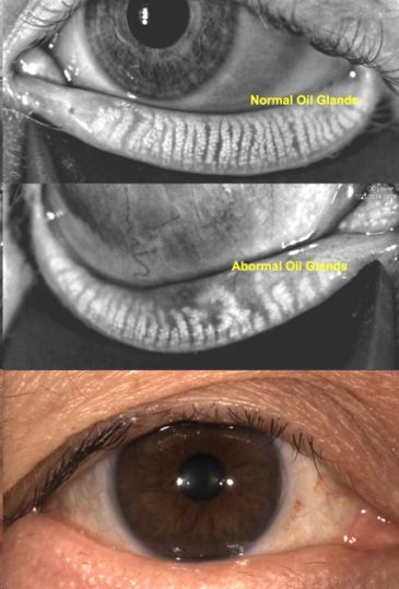
A major cause of chronic dry eye disease is Meibomian Gland Dysfunction (MGD).
Infrared imaging reveals damage and loss of the eyelid's oil-producing glands that may not be visible during a traditional eye examination.
Progressive gland loss can be irreversible, contributing to chronic inflammation, eye discomfort and fluctuating vision that can affect daily activities and work.

Seeing 20/20 doesn't always mean your eyes and brain work well together.
Many children and adults have difficulties with eye coordination, focusing, tracking or visual processing that may go undetected during routine eye examinations.
These difficulties can affect reading, learning, work, sports and everyday activities. They may also devel
Seeing 20/20 doesn't always mean your eyes and brain work well together.
Many children and adults have difficulties with eye coordination, focusing, tracking or visual processing that may go undetected during routine eye examinations.
These difficulties can affect reading, learning, work, sports and everyday activities. They may also develop following a concussion, stroke or other neurological conditions.
Our clinic provides specialized functional vision assessments and individualized vision therapy to help patients develop or regain visual skills, improve visual comfort and reach their potential.

Children often have eye health or vision problems without noticeable symptoms and may not recognize or be able to explain what they are experiencing. Clear vision is only part of healthy eye development.
Optomap® ultra-widefield retinal imaging helps detect retinal abnormalities that may otherwise go unnoticed, while axial length measureme
Children often have eye health or vision problems without noticeable symptoms and may not recognize or be able to explain what they are experiencing. Clear vision is only part of healthy eye development.
Optomap® ultra-widefield retinal imaging helps detect retinal abnormalities that may otherwise go unnoticed, while axial length measurements monitor eye growth and myopia progression to guide early management.
When indicated, specialized binocular and functional vision assessments help identify difficulties with eye coordination, focusing and tracking that may affect learning and daily activities.
When we're young, we rarely think about eye disease, especially if we see clearly. Yet retinal holes or tears can occur at any age or prescription, particularly with high myopia.
Optomap® retinal imaging helps detect retinal abnormalities, while dry eye assessments and meibography can identify chronic dry eye problems affecting daily comfo
When we're young, we rarely think about eye disease, especially if we see clearly. Yet retinal holes or tears can occur at any age or prescription, particularly with high myopia.
Optomap® retinal imaging helps detect retinal abnormalities, while dry eye assessments and meibography can identify chronic dry eye problems affecting daily comfort and work. Sometimes, difficulties with reading or computer work arise from dry eye or undetected binocular vision problems, rather than an incorrect glasses prescription.
As we approach middle age, OCT macular and optic nerve scans can help detect early changes associated with myopic macular disease, epiretinal membranes and glaucoma, sometimes before noticeable vision loss.
As we age, macular diseases become increasingly common, often without noticeable symptoms. Conditions such as age-related macular degeneration (AMD), epiretinal membranes and myopic macular degeneration may be missed during routine examinations.
OCT macular scans, especially after age 60–65, combined with Optomap® retinal imaging, help det
As we age, macular diseases become increasingly common, often without noticeable symptoms. Conditions such as age-related macular degeneration (AMD), epiretinal membranes and myopic macular degeneration may be missed during routine examinations.
OCT macular scans, especially after age 60–65, combined with Optomap® retinal imaging, help detect and monitor subtle changes early, when timely care may prevent permanent vision loss. Optic nerve OCT also helps assess glaucoma risk.
Dry eye and meibomian gland dysfunction (MGD) assessments are especially important before cataract surgery, particularly with specialty IOLs, to improve measurement accuracy and optimize visual outcomes.
Diabetes can damage the retina and macula without noticeable symptoms.
Optomap® retinal imaging and OCT macular scans help detect and monitor diabetic retinopathy and macular swelling.
Arthritis and autoimmune conditions can cause dry eye, uveitis and other eye inflammation. Certain medications also carry risks: Plaquenil® may cause retina
Diabetes can damage the retina and macula without noticeable symptoms.
Optomap® retinal imaging and OCT macular scans help detect and monitor diabetic retinopathy and macular swelling.
Arthritis and autoimmune conditions can cause dry eye, uveitis and other eye inflammation. Certain medications also carry risks: Plaquenil® may cause retinal toxicity, long-term steroids can increase glaucoma and cataract risk, and Accutane® may contribute to dry eye and contact lens discomfort.
Appropriate retinal imaging, OCT and dry eye assessments help our doctors identify potential complications early and personalize your eye care.